


Osteochondrosis is a disease of the spine, characterized by degeneration of the intervertebral disc with a significant reduction in its height, sclerosis of the disc surfaces of the vertebrae and the reactive growth of regional osteophytes.
The cartilage tissue of the discs affected by osteochondrosis is gradually reborn and becomes a similarity to bone.The hardened disc decreases in size, loses the properties of the shock absorber between the vertebrae and begins to press on the nerve endings, leading to the appearance of painful sensations.
The initial stage of osteochondrosis most often does not manifest with unpleasant sensations in the spine and can be diagnosed as a disease of the internal organs and a true diagnosis is detected only after multiple examinations pass.
The cervical, thoracic, lumbar, sacral and ordinary osteochondrosis are distinguished through localization.Lumbar osteochondrosis (over 50%of cases), cervical osteochondrosis (more than 25%) and total (about 12%) are most commonly diagnosed.
The intervertebral disc is a fiber plate.In the middle of the disc there is a nucleus surrounded by a fibrous ring (a cloth that resembles tendons).The intervertebral disc does not have its vascular system and therefore eats at the expense of other fabrics.An important source of nutrients for the disc is the posterior muscles, it is their dystrophy that most often leads to the development of the disease.When you lift weights, jumps and other physical exertion, the discs act as a shock absorber and maintain the necessary distance between the vertebrae.As the largest load falls on the lumbar spine, it is in it, the convexity and the intervertebral hernias, which are a complication of this disease, are most commonly formed.
Protection of the intervertebral disc- Convexity (prolapse) of the disc without rupture of the fibrous ring.
Hernia on the intervertebral disc- convexity (prolapse) of a disc with a tearing of a fibrous ring and a "flow" of a reactive nucleus.Especially often the hernia is formed when the spine is formed or during the simultaneous slope and rotation of the torso sideways, especially if there is a heavy object in the hands.In this position, the intervertebral discs experience a very high load, the pressure inside the intervertebral disc rises, the vertebrae is pressed on one side of the disc, and the core is forced to shift to the opposite side and press on the fibrous ring.At some point, the fibrous ring does not withstand such loading and the disc is protruding (the fibrous ring is stretched but remains whole) or the hernia is formed (the fibrous ring breaks and part of the contents of the nucleus is "flowing" through the breakthrough).With the increase in the spinal cord load and the creation of conditions for increasing the pressure in the damaged intervertebral discs, the hernia increases in size.
This is very important for the direction of convexity and the size of the hernia, if the hernia comes forward or on the side, it can lead to pain and impaired work of some organs and when it sticks out to the spinal cord and damage, the consequences can be much more serious.If the intervertebral hernia affects the nerve processes or the roots of a particular segment of the spine, this leads to a violation of the organ function for which the damaged segment of the spine is responsible.Another option is possible: due to the bulge of the disc in one direction, in the opposite side the distance between the vertebrae decreases and this leads to a pinch of nerve processes from the vertebrae themselves.The intervertebral hernia in the lumbar region most often causes pain in the legs, hernias in the thoracic region contributes to dizziness, heart pain, respiratory disorders, etc., hernia in the cervical spine can cause headaches, dizziness and numbness of the hands.The most dangerous intervertebral hernias are more than 10 mm in size, a sharp narrowing of the spinal canal, tightening blood vessels and injuring nerve endings, prolonged exposure, which leads not only to severe pain but also to blood disorders, loss of sensitivity in the limbs with subsequent complications.But the most dangerous are the sequestrated hernias of the intervertebral discs, ie.Hernia is ready to destroy or separate the fragment, followed by its descent into the spinal canal, which can have severe consequences: impaired functions of the pelvic organs and paralysis of the lower limbs.
Causes of osteochondrosis
The causes that cause changes in the intervertebral discs are not fully studied.People begin to experience osteochondrosis most often after 35 years.The development and exacerbation of this disease is facilitated by various back injuries, static and dynamic overloads, as well as vibrations.The more adult a person, the more manifestations there are.But in recent years, more and more people aged 18 to 30 have been complaining of back pain.There are many reasons for the early manifestation of the disease: weak physical exercise, disorder of the posture and curvature of the spine, flat legs and excess weight.
So, we will emphasize the main reasons:
- hereditary (genetic) predisposition;
- metabolic disorder in the body, infection, intoxication;
- Overweight, malnutrition (lack of trace elements and fluid);
- Age -related changes;
- spinal injuries (bruises, fractures);
- Stand disturbance, spinal curvature, hypermobility (instability) of the spine segments, flat legs;
- adverse environmental conditions;
- A sedentary lifestyle;
- work associated with lifting weights, frequent changes in body position (turns, bending and expansion, rattling movements);
- prolonged exposure to uncomfortable postures in an upright position, sitting, lies, when lifting and transferring weights, when performing other work in which the pressure in the discs and the load on the spine as a whole increases;
- excessive physical activity, unevenly developed muscle system;
- Spinal overloads associated with foot diseases, as well as as a result of wearing uncomfortable shoes, high heels and pregnancy in women;
- a sharp termination of regular training from professional athletes;
- nerve overvoltage, stressful situations, smoking;
- Hypothermia, adverse weather conditions (high humidity at low temperature).
Symptoms characteristic of osteochondrosis
Patients suffering from osteochondrosis complain of constant back pain, to which numbness and feeling of limb pain often join.In the absence of adequate treatment, weight loss and atrophy of the limbs occurs.
The main symptoms:
- constant diseased pain at the back, feeling of tingling and limb pain;
- increased pain during sudden movements, exercise, weight lifting, coughing and sneezing;
- reduction in the volume of movement, muscle cramps;
- With osteochondrosis of the cervical spine: pain in the hands, shoulders, headache;It is possible to develop the so -called spinal artery syndrome, which consists of the following complaints: noise in the head, dizziness, flies of "flies", colored spots in front of the eyes in combination with a burning pulsating headache.The cause of the spinal artery syndrome may be its spasm in response to the direct irritation of its sympathetic split due to bone growth, disc herniation, arthrosis of the intervertebral joint and the reflex reaction due to irritation to any receptors of the spine.The presence of spinal artery syndrome can worsen the course of coronary or cardiac-muscular pathology, if any;
- With osteochondrosis of the chest: chest pain (as a "bet" in the chest), in the heart and other internal organs;
- With osteochondrosis of the lumbosacral spine: lower back pain, radiant in the sacrum, lower limbs, sometimes in the pelvic organs;
- The lesion of nerve roots (with hernias of the intervertebral discs, bone growths, spondylolist, spondylo arthrosis): pain in shooting and sensitivity, hypotrophy, hypotension, weakness in innervized muscles and reduction of reflexes.
Diagnosis of osteochondrosis
The preliminary diagnosis was established during the patient's initial examination.The study is usually conducted by a neurologist in connection with the patient's complaints about local changes, which can be manifested by pain, deformation or restriction of mobility.The spine is examined in the position of the patient standing, sitting and lying, both at rest and on the move.The level of spinal damage is determined by counting the number of vertebrae from certain anatomical landmarks or according to a special scheme.
When the back is examined, the attention is drawn to the posture, the structural characteristics of the body, the line of the spinous processes (medium groove), the lower corners of the shoulders, the ridges of the iliac bones, the side contours of the waist and neck, the rejection of the interior of the furrows are paid, the rejection of verticals are paid.
The sensation of the spine allows you to supplement the examination data (presence or absence of deformity), to determine the location, extent and nature of the pain.When you feel, the tension of the muscles located next to the spine is also noted, since most injuries and diseases of the spine are accompanied by an increase in muscle tone.
Spinal flexion is used to determine the amplitude of movements in different parts of the spine.
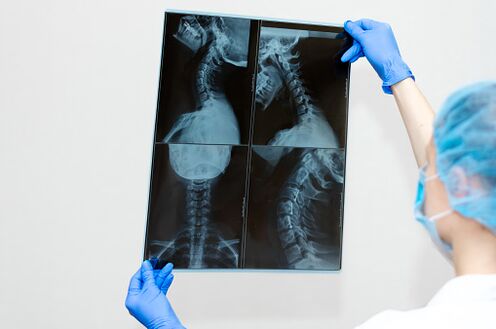
The main role in the study of the spine is assigned to radiography, computed tomography and magnetic resonance imaging, which determines the level of damage, the diagnosis and betrayal, hidden pathologies are revealed.These diagnostics allow the attending physician to determine the treatment tactics and to choose the most effective treatments.
Methods of treating osteochondrosis and its complications
Treatment of osteochondrosisAnd its complications are performed using conservative methods aimed at eliminating pain syndrome, disorders of spinal roots and preventing the progression of dystrophic changes in the structures of the spine.With the ineffectiveness of conservative treatment and in special indications, surgically (surgical) treatment is performed whose volume depends on the level of damage and clinical manifestations of the disease.
The duration of treatment of osteochondrosis and its complications depends mainly on the severity of the disease, age -related changes, treatment methods, as well as on the conscientious prescription and recommendations of the treating physician.As practice shows, the active phase of treatment in most cases lasts 1-3 months using conservative methods, and the recovery period after surgery is about 1 year.At the onset of treatment in some patients, it is possible to increase the pain syndrome associated with the reaction of the muscle system and other entities to expose unusual to the body.The pain stops for a short time with physiotherapy procedures, medicines, and special exercise.The result of treatment largely depends on the behavior of the patients themselves, which requires patience, perseverance, perseverance, a certain force of will, as well as the desire for recovery.The largest efficiency of conservative therapy and rehabilitation after surgery can be achieved in conditions of specialized medical centers and sanatoriums, equipped with a modern diagnostic and therapeutic base, as well as highly qualified practitioners who use the complete treatment of diseases of the musculoskeletal system.
Complex conservative treatment includes physiotherapy, physiotherapy, massage, manual therapy, grip (grip) of the spine, reflexology and drug therapy.
Medical Physical Education (Exercise Therapy)-The main method of conservative treatment of diseases of the musculoskeletal system is to create dosage loads aimed at decompression of nerve roots, correction and strengthening of the muscle corset, increase in volume and development of the necessary movement and the proper lifeline.This is achieved through regular classes in rehabilitation equipment and joint gymnastics.As a result of the exercises, blood circulation improves, metabolism and nutrition of the intervertebral discs are normalized, the intervertebral space increases, a muscle corset is formed and the load on the spine is reduced.
Physiotherapy is a method of treatment that uses physical factors: low frequency currents, magnetic fields, ultrasound, laser and more.It is used to relieve pain, inflammatory processes, rehabilitation after injuries and surgery.When the methods of physiotherapy are used, the treatment of many diseases is reduced, the effectiveness of the use of drugs and their dose reduction increases, there are no side effects inherent in drug treatment.
Massage is a set of techniques of mechanical dosage effects in the form of friction, pressure, vibrations performed directly on the surface of the human body with hands.Effectively relieves muscle tension, muscle pain, improves blood circulation, has a common strengthening effect.
Manual therapy is an individually selected manual effect on the bone system -a muscle system for the removal of acute and chronic pain in the spine and joints, as well as an increase in the volume of movement and adjustment of the posture.One of the areas of manual therapy is visceral manual therapy, which helps to restore normal organs mobility, improves blood supply, lymphocycling, normalizes metabolism, restores immunity, prevents the spread of chronic diseases.
The enlargement (grip) of the spine is an effective method of treating spinal pain syndromes and joints using individually selected load using special equipment.The procedure is aimed at increasing the intervertebral space, eliminating pain and restoring an anatomically correct spinal shape.
Reflexology - various therapeutic techniques and methods for influencing the reflexogenic zones of the human body and acupuncture points.The use of reflexology in combination with other therapeutic methods significantly increases their effectiveness.Most often, reflexology is used for osteochondrosis, accompanied by pain, nervous system diseases, sleep disorders, mental imbalance, and overweight and tobacco curls.By acting at certain points, one can bring the body into harmony and treat many diseases.
Drug therapy is indicated during the period of exacerbation of the disease aimed at stopping the pain syndrome, eliminating the inflammatory process and increasing the metabolic processes by taking or administration of drugs with the help of intramuscular or intravenous injections.
Although each of the above methods is a highly effective, still sustainable therapeutic effect can only be obtained with their combination with classes on rehabilitation equipment, ie.When creating a full muscle corset.